
Panoramic X-Ray Unit in Burnaby
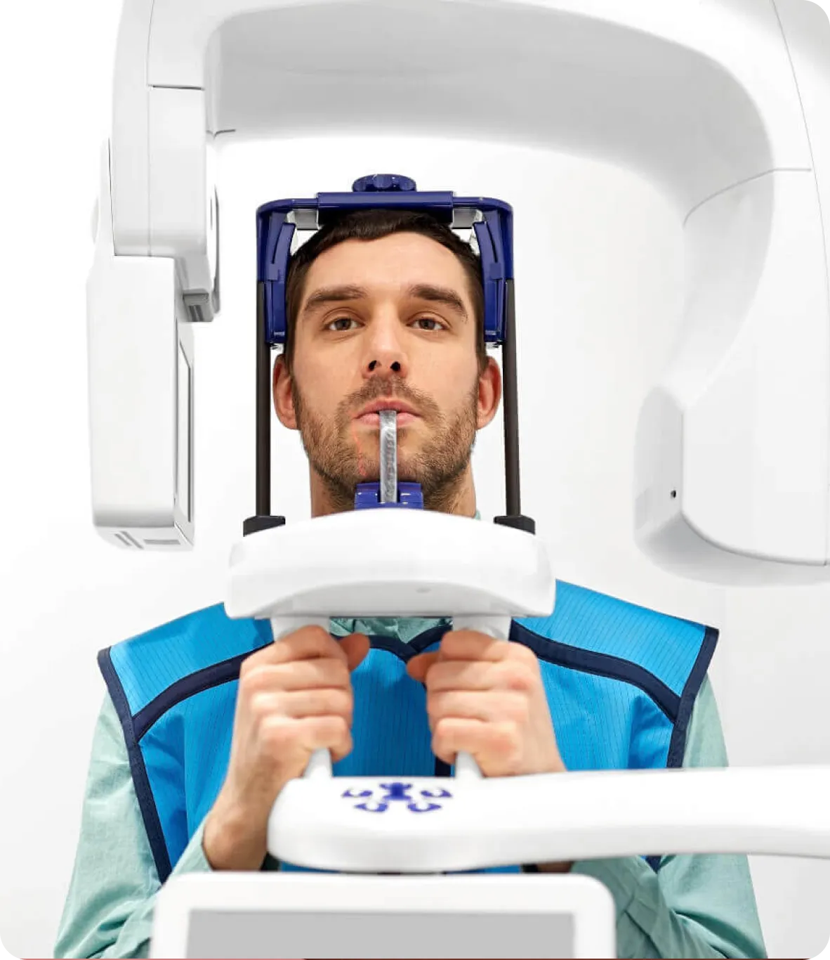
Panoramic radiography uses a rotating X-ray unit to create a broad two-dimensional image of the teeth, jaws, and selected surrounding structures. At Burnaby Dentist & Orthodontist, panoramic imaging is used when clinically indicated to support assessment and treatment planning.

What our Patient Think About Us?
Posted on Google loveleen dhanoa
Trustindex verifies that the original source of the review is Google.
I was scheduled for full checkup and cleaning. Randeep was so sweet, polite and experienced. At every point of the appointment, she made sure that I knew what was happening and If I was in any pain. She made my experience very smooth and less scary. She did an amazing job on cleaning and explaining everything. Dr. Sasan explained clearly about what is needed or not about my dental insecurities. Front Staff was nice and polite. Highly recommended!Posted on Google James YipAmazing staff and facilities here and had a great time doing regular check up and cleanings. Would love to be backPosted on Google
 fardad faridiThanks for your good servicePosted on Google
 Alyoshka MyshkoPosted on Google
 Carlotta CrestanelloThe best experience!! I was so nervous for my wisdom tooth extraction and the whole staff was so supportive. Highly recommended, everyone should feel like this when they go to the dentist. I ended up having fun!!!Posted on Google
 Sunny Lee-GordonGhazal was informative and caring! Loved my cleaning session with her :) definitely will come back!Posted on Google
 Elan AsselstineHave come here for several years and now come with my kids (7 and 4), great experience, very friendly.Posted on Google
 Andrew WuPatient and kind with their service. Much needed when it comes to teeth cleaningPosted on Google
 Layla SongThe dentist was professional and gentle – the whole experience was smooth and comfortable. Highly recommend!Posted on Google
 Jas JaskaranGreat service, staff and doctor are friendly. They’re giving very open and transparent service so feel comfortable with all the procedures and treatments.
What Is a Panoramic Dental X-Ray?
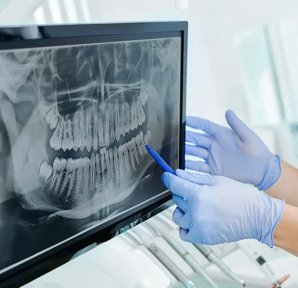
A panoramic dental X-ray, also known as a panoramic radiograph or orthopantomogram (OPG), is a two-dimensional imaging technique that provides a broad view of the teeth, jaws, and selected surrounding structures in a single image. It allows the dentist to evaluate overall relationships across the mouth as part of a comprehensive clinical assessment.

How Panoramic Imaging Works
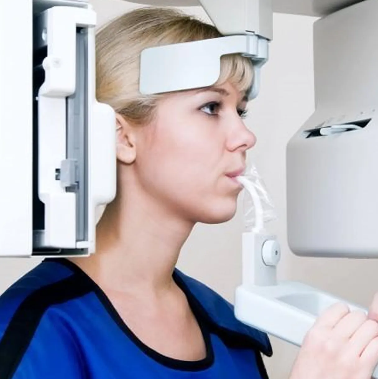
A panoramic X-ray uses a rotating machine that moves around your head to take a two-dimensional panoramic image of your mouth. You simply remain still while the rotating unit captures the image. The exact exposure time depends on the equipment and imaging program being used. The result is a digital image that provides a broad view of the teeth, jaws, and selected surrounding structures.

What Areas Are Captured in One Image
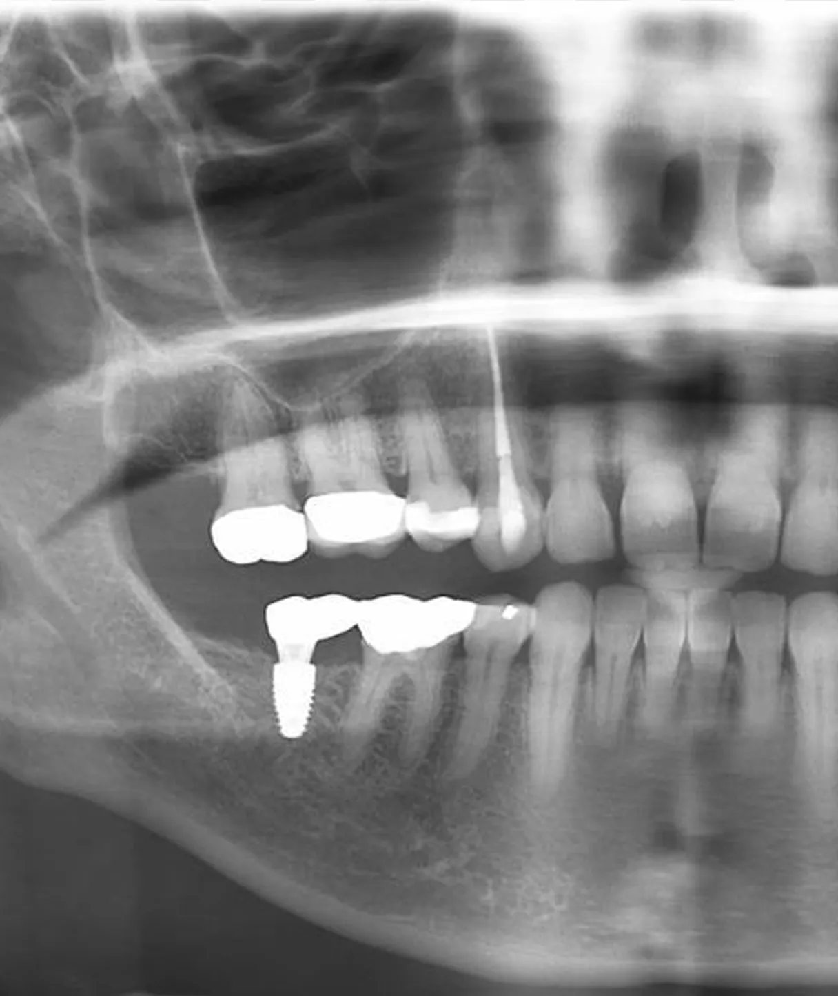
A single panoramic X-ray provides a broad view of the upper and lower teeth, jawbones (maxilla and mandible), sinuses, temporomandibular joints (TMJ), and surrounding oral and facial structures in one image.

How It Differs From Traditional Bitewing X-Rays
Bitewing X-rays focus on small, detailed sections of the mouth, typically used to detect cavities between teeth. In contrast, a panoramic X-ray provides a full-mouth overview in one scan, showing broader structures and relationships.
Both types of imaging are often used together to provide a broader understanding of your oral health.
At Burnaby Dentist and Orthodontist Clinic
Begin Your Smile Journey with Dr. Hadianfar

Why Dentists Use Panoramic X-Rays
Here are the key reasons dentists rely on panoramic X-rays to support diagnosis and patient care.

Viewing Broad Oral and Jaw Structures
A panoramic radiograph shows the upper and lower dentition, jawbones, and selected surrounding structures in one broad image. It helps the dentist assess general development, tooth position, jaw relationships, and other findings that may require closer investigation.

Detecting Issues Below the Surface
Panoramic images may show radiographic findings that are not visible during a direct examination, such as impacted teeth, altered eruption, certain areas of bone loss, jaw changes, or larger areas of infection or pathology. Findings are interpreted together with symptoms, clinical examination, and any additional imaging that may be required.

Supporting Timely Assessment and Planning
When a panoramic radiograph is clinically indicated, it may help document developmental, structural, or pathological findings that require further assessment. The dentist uses the image with the rest of the examination to decide whether monitoring, additional imaging, referral, or treatment should be considered.

Supporting Timely Assessment and Planning
When a panoramic radiograph is clinically indicated, it may help document developmental, structural, or pathological findings that require further assessment. The dentist uses the image with the rest of the examination to decide whether monitoring, additional imaging, referral, or treatment should be considered.
What Can a Panoramic X-Ray Help Evaluate?
A panoramic radiograph can help the dentist evaluate broad patterns and structures across the jaws. It may reveal findings that warrant monitoring, targeted dental X-rays, specialist assessment, or other diagnostic testing.

Impacted or Missing Teeth
Panoramic images may help assess tooth development, eruption patterns, impacted wisdom teeth, unerupted teeth, and whether expected teeth are visible within the jaws. Clinical records and other imaging may be needed to determine whether a tooth is congenitally absent, previously removed, or otherwise not visible.
Jawbone and Visible Sinus Areas
A panoramic radiograph may show broad changes in the upper and lower jawbones and portions of the maxillary sinuses that appear within the image. Findings in these areas may require targeted dental imaging, cone-beam CT, medical imaging, or referral for further evaluation.
General Jaw-Joint and Skeletal Findings
A panoramic image may show the general shape and bony appearance of the jaw joints and the relationship of the jaws and teeth. It does not fully assess joint movement, muscles, the articular disc, or all causes of temporomandibular disorders. Additional clinical evaluation or other imaging may be recommended when TMJ or TMD symptoms are present.
Areas Requiring Further Evaluation
Panoramic images may show areas of altered bone density, radiolucency, radiopacity, or changes around tooth roots that warrant closer evaluation. These findings do not establish a diagnosis on their own and may require targeted imaging, follow-up, referral, or laboratory assessment.
Panoramic X-Rays vs Traditional Dental X-Rays
Panoramic and traditional dental X-rays each play an important role in providing a complete picture of your oral health. While they are different in scope and detail, they are often used together to aid clinical evaluation.

Full-View Imaging vs Close-Up Detail
A panoramic X-ray offers a broad, full-mouth view in a single image, showing the teeth, jaws, and surrounding structures. In contrast, bitewing and periapical X-rays focus on smaller, specific areas to capture fine detail of individual teeth and nearby bone.

When Each Type Is Used
Different X-rays are used depending on the situation. Routine checkups may include either type based on clinical need, while emergency visits often require targeted imaging. Panoramic X-rays are especially useful in orthodontic assessments and overall dental assessment.

How They Work Together for Diagnosis
These imaging methods are complementary rather than interchangeable. Together, they give dentists both a wide overview and detailed insight, providing a broader view alongside detailed imaging of oral health.

Limitations of Panoramic Imaging
A panoramic X-ray provides a broad overview rather than fine detail of every tooth. Because the curved jaws are represented in a flat two-dimensional image, some areas may appear enlarged, overlapped, distorted, or less sharply focused. Bitewing, periapical, cephalometric, cone-beam CT, or other imaging may be recommended when more specific information is needed.
Schedule a Panoramic X-Ray Consultation
Schedule a dental examination to learn whether panoramic imaging may be clinically appropriate. We’re here to help you feel informed, comfortable, and confident about your care.
What Is the Panoramic X-Ray Process Like?
Panoramic imaging is performed outside the mouth, although a small bite guide may be used to help position the teeth. The dental team explains the steps, adjusts the unit where possible, and asks you to remain still while the image is captured.

Standing or Sitting Positioning
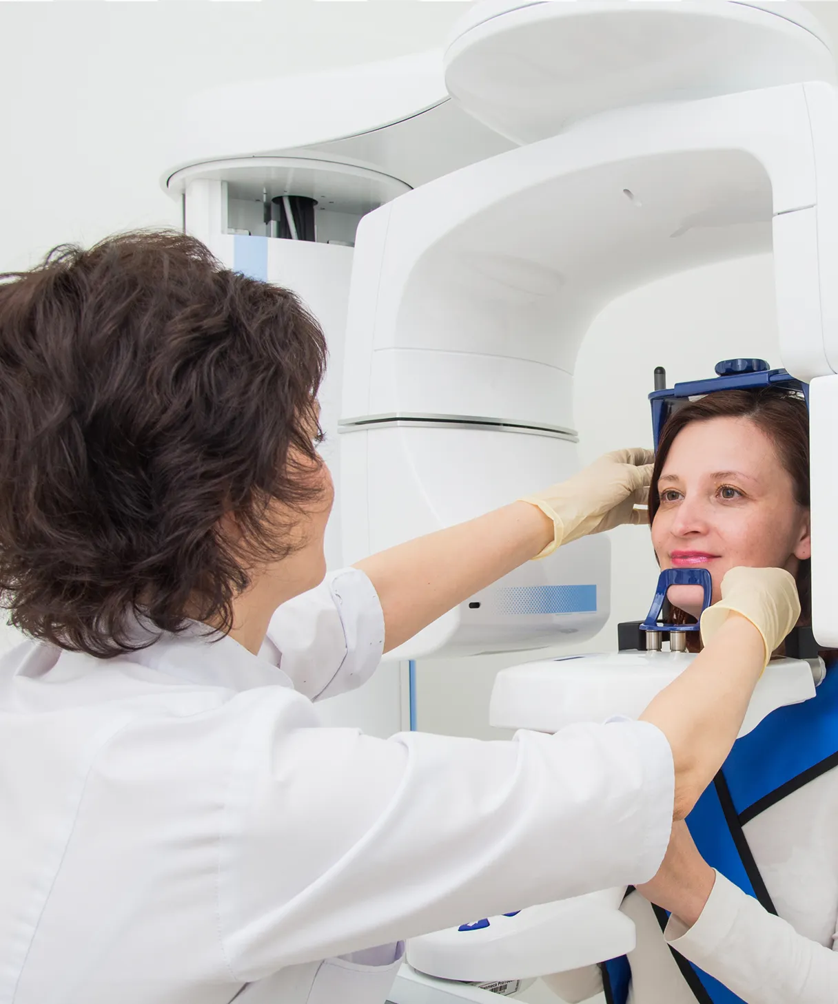
We will position you comfortably while either standing or sitting, depending on what is best for you. Your head is gently stabilized to ensure clear and diagnostically useful imaging while you remain relaxed and still.
Brief Image Capture
Once you are positioned, the rotating arm usually takes only a short time to travel around your head. The exact exposure time depends on the panoramic unit and imaging program. Remaining still and following the positioning instructions helps reduce motion blur and the need for retakes.
What the Positioning May Feel Like
During the scan, you may be asked to bite gently on a positioning guide while your head is supported and to remain still as the machine rotates around you. Most patients tolerate the process well, although positioning may be less comfortable for those with jaw pain or limited mobility.
What the Positioning May Feel Like
During the scan, you may be asked to bite gently on a positioning guide while your head is supported and to remain still as the machine rotates around you. Most patients tolerate the process well, although positioning may be less comfortable for those with jaw pain or limited mobility.
Is Panoramic X-Ray Imaging Safe?
Panoramic radiography uses ionizing radiation. The dose from a properly performed dental X-ray is generally low, but each image should still be clinically justified and taken using settings appropriate to the patient and diagnostic purpose.
Clinically Justified Exposure
A panoramic X-ray is recommended only when the expected diagnostic information is relevant to the patient’s examination, symptoms, development, or treatment planning. The exposure settings are selected to obtain a diagnostically useful image while limiting radiation as much as reasonably achievable.
Optimized Digital Imaging
Digital panoramic systems allow images to be viewed electronically after exposure. Image quality and radiation dose depend on the equipment, patient positioning, selected program, and exposure settings. The unit should be maintained and operated according to applicable quality-assurance and radiation-protection requirements.
Positioning and Exposure Measures
Radiation protection includes prescribing the image only when clinically justified, selecting settings appropriate to the patient and imaging purpose, positioning the patient carefully, observing the patient during exposure, and avoiding unnecessary retakes. Protective shielding is used when appropriate and when it will not interfere with the required image.
How Often Is a Panoramic X-Ray Needed?
We recommend panoramic X-rays only when they are clinically necessary, based on your individual oral health and treatment needs. They are not required at every visit and are used thoughtfully as part of your overall care.


Based on Individual Dental Needs
There is no standard schedule that applies to every patient. The dentist considers your age, development, symptoms, oral-health history, previous images, examination findings, and planned treatment before recommending another panoramic radiograph.
Use During New-Patient Assessment
A panoramic X-ray may be recommended during a comprehensive dental exam when your health history, clinical examination, symptoms, previous records, or planned treatment indicate that a broad view of the teeth and jaws would provide useful diagnostic information. It is not routinely required for every new patient.
Monitoring Changes Over Time
A repeat panoramic image may be considered when it is expected to provide clinically useful information about development, healing, disease progression, or treatment. Existing images should be reviewed before additional exposure is prescribed.
Does a Panoramic X-Ray Replace Other Dental Imaging?
A panoramic X-ray is a valuable diagnostic tool, but it does not replace all other forms of dental imaging. It provides a broad overview of the mouth, while other imaging methods are often needed for more detailed assessment.

When Additional X-Rays Are Required
Additional X-rays are often needed for detailed cavity detection and close-up evaluation of individual teeth, sometimes alongside tools such as an intraoral camera to provide more detailed visual assessment. Focused imaging allows dentists to see fine structural details that may not be visible in a panoramic scan.

Combining Imaging for Complete Assessment
Different radiographic views answer different clinical questions. Bitewing radiographs may provide more detail for certain areas between teeth, while periapical images may show individual teeth and surrounding bone more closely. The dentist selects only the images needed for the particular examination or treatment plan.

Personalized Diagnostic Planning
The choice of imaging is always based on individual patient needs. Dentists select the most appropriate combination of X-rays to support assessment of oral health.
CDCP
Important Information for CDCP Patients
Treatment under the Canadian Dental Care Plan is not fully covered. A co-payment will apply.

Why Burnaby Patients Trust Us for Dental Imaging
Burnaby Dentist & Orthodontist provides dental imaging services with a focus on careful assessment, patient safety, and clear communication. Every panoramic X-ray is performed carefully to support clinical assessment and treatment planning.
Modern, High-Quality Imaging Technology
Panoramic and other digital X-ray systems provide different types of information for clinical assessment. The appropriate image is selected according to the area being evaluated and the diagnostic question that needs to be answered.
Careful Use of Diagnostic X-Rays
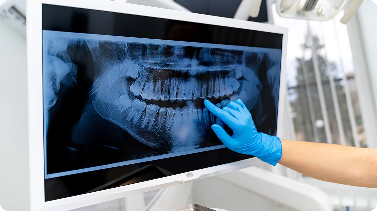
Dental X-rays are recommended according to individual clinical need rather than taken automatically at every visit. Previous images, examination findings, symptoms, development, and planned treatment are considered before additional imaging is prescribed.
Clear Explanations of Findings
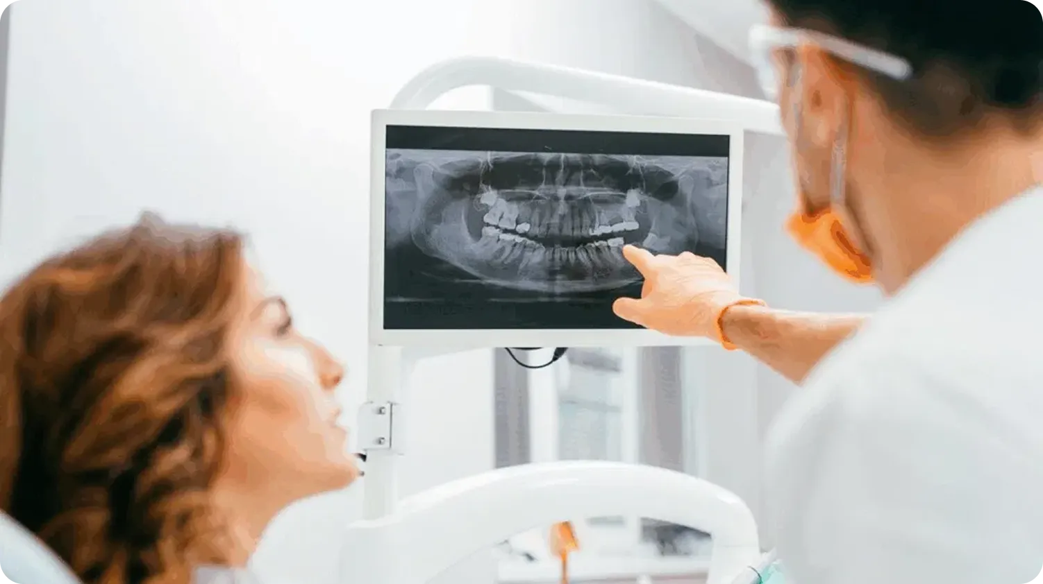
The dentist can review relevant radiographic findings with you and explain whether monitoring, additional imaging, referral, or treatment may be considered. You can ask questions before making decisions about care.
Focus on Safety & Preventive Care
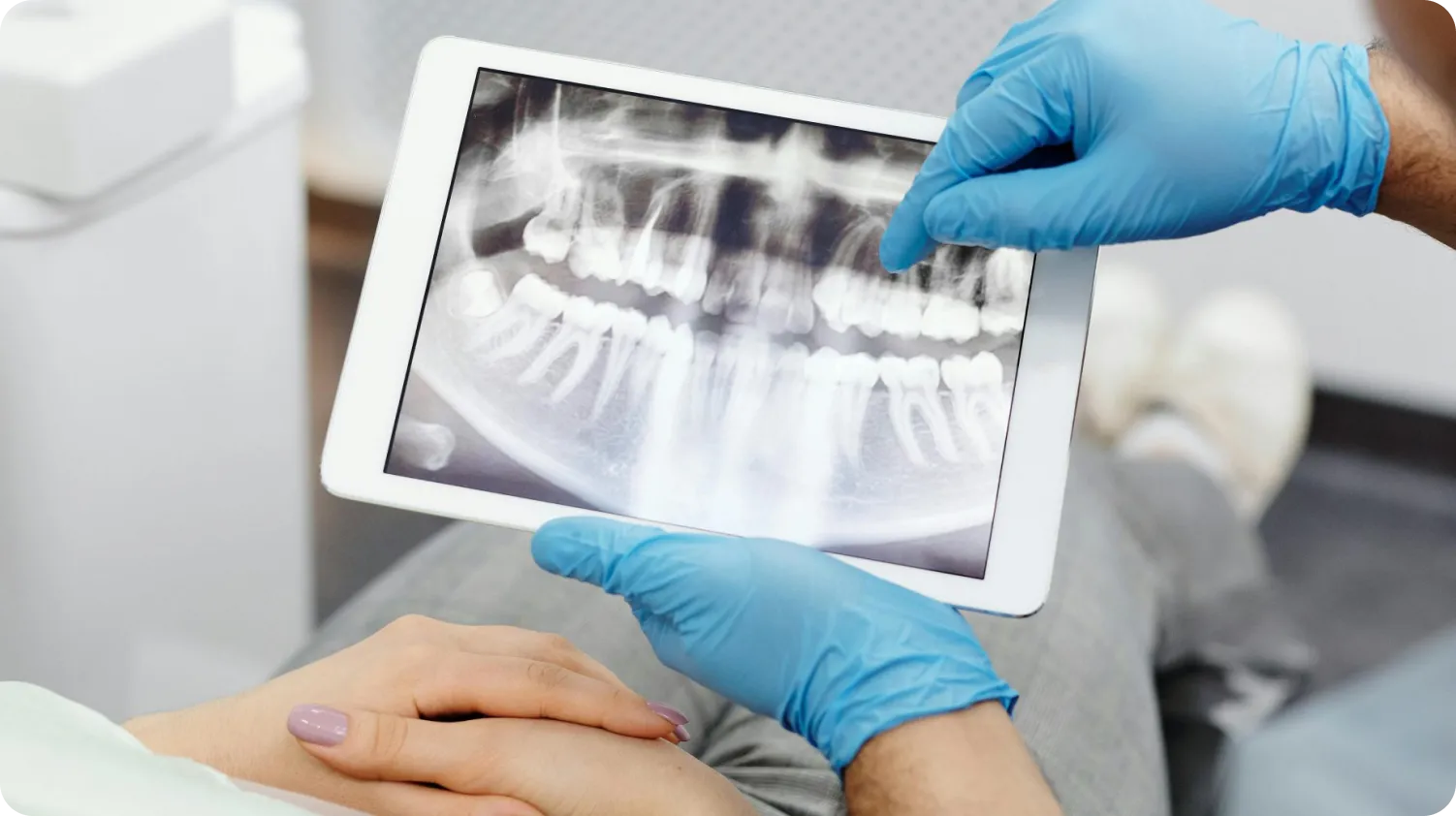
Panoramic images are taken using appropriate positioning, exposure settings, and quality-control procedures. Avoiding unnecessary images and retakes is an important part of limiting radiation exposure.
Plan Your Visit to Burnaby Dentist and Orthodontist
If you are experiencing tooth pain or a dental concern, you can contact Burnaby Dentist and Orthodontist to request an appointment and discuss your situation with our team. Our clinic is located at 3999 Kingsway, Burnaby, BC V5H 1Y8, serving patients from Metrotown, South Burnaby, and nearby communities.
Same-day emergency appointments may be available depending on scheduling, though availability and treatment timing cannot be guaranteed. Please contact us to check availability and arrange your visit.
Our team can help you understand your symptoms and guide you through appropriate next steps based on your individual needs.
FAQs
Why would I need a panoramic X-ray?

It may be recommended when the dentist needs a broad view of the teeth, jaws, and surrounding structures to support clinical assessment and treatment planning.
What does a panoramic X-ray show?
It shows all teeth, upper and lower jaws, sinuses, jaw joints (TMJ), and surrounding bone structures.
Does a panoramic X-ray hurt?
The X-ray exposure itself does not cause a physical sensation. You may feel light pressure from the chin rest, head supports, or bite guide used for positioning. Let the dental team know if you have jaw pain, limited mobility, or difficulty maintaining the required position.
How long does a panoramic X-ray take?
Positioning usually takes longer than the exposure itself. The image capture generally lasts only several seconds, although the exact time varies by the equipment and imaging protocol.
How often should I get a panoramic X-ray?
It depends on individual dental needs and is not required at every visit.
Contact Us Today!
Contact Burnaby Dentist and Orthodontist About Panoramic X-Ray in Burnaby
- Straighten crooked and misaligned teeth
- Conceal chips, cracks, and discoloured teeth
- Whiten and brighten your smile
- Take years off the look of your teeth
- Boost your self-esteem!
Monday
9 AM – 5 PM
Tuesday
8 AM – 5 PM
Wednesday
9 AM – 6 PM
Thursday
9 AM – 5 PM
Friday
9 AM – 5 PM
Saturday
9 AM – 4 PM